Your eyes can sometimes develop problems without causing noticeable symptoms in the early stages. This is why detailed eye examinations can be important, particularly for people with diabetes, glaucoma, retinal conditions, or unexplained changes in vision. An OCT scan is a non-invasive imaging test that provides detailed images of the retina and helps eye doctors examine the different layers of the eye. A Retina Specialist Doctor in Gunjur can recommend an OCT scan when a closer evaluation of the retina is needed.
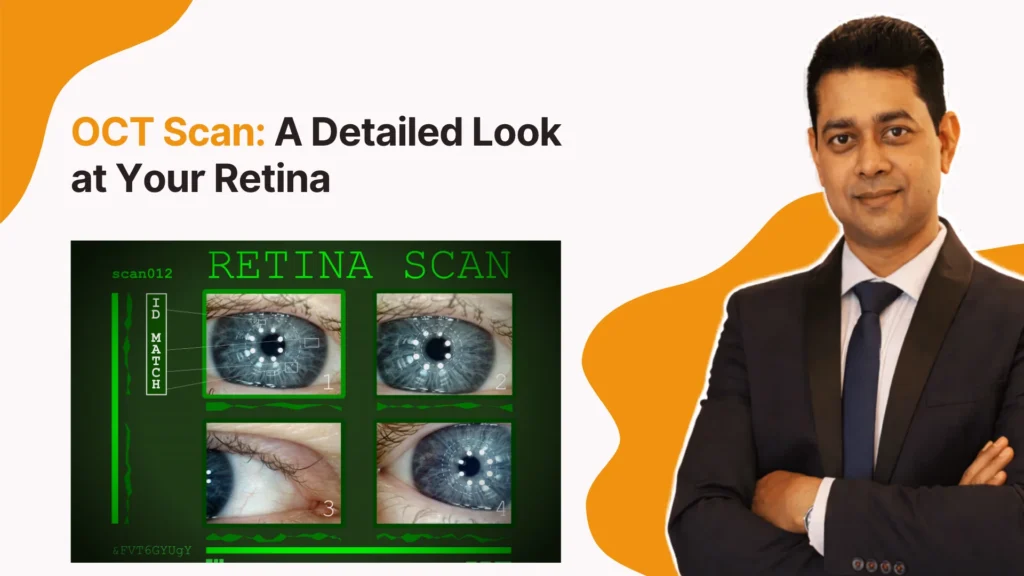
In this video, the doctor explains what an OCT scan is, how it helps examine the retina, and why this detailed eye imaging may be recommended for certain patients.
OCT stands for Optical Coherence Tomography. It is an advanced imaging technique that uses light to create detailed cross-sectional images of the retina.
Unlike a routine eye examination, which allows the doctor to examine the eye from different perspectives, OCT can show the individual layers of the retina. This can help doctors identify subtle changes that may not be obvious during a basic examination.
The test is non-invasive and generally quick to perform.
The retina is a light-sensitive layer at the back of the eye that plays an important role in vision. Conditions affecting the retina can sometimes progress gradually.
An OCT scan can help doctors examine the retinal structure and identify changes such as swelling, thinning, or fluid accumulation. It can also help monitor changes over time and assess how the retina responds to treatment.
An eye doctor may recommend OCT imaging when evaluating or monitoring conditions such as:
- Diabetic eye disease
- Macular degeneration
- Macular oedema
- Glaucoma
- Retinal swelling
- Macular holes
- Epiretinal membranes
- Unexplained changes in vision
The need for an OCT scan depends on the patient's symptoms, examination findings, age, medical history, and risk factors.
An OCT examination usually does not require direct contact with the eye. The patient sits in front of the imaging device and looks at a target while the machine captures images.
The scan itself is generally quick and painless. In some cases, the doctor may use dilating eye drops before the examination to obtain a more detailed view of the retina.
Your eye specialist will explain whether dilation is required based on the examination.
No. OCT is a valuable diagnostic tool, but it is only one part of a comprehensive eye assessment.
An eye doctor may combine OCT findings with visual acuity testing, eye pressure measurement, retinal examination, medical history, and other investigations to understand the patient's condition.
This combination helps the doctor interpret the scan in the correct clinical context.
Changes such as blurred vision, distortion, new floaters, flashes of light, or sudden loss of vision should not be ignored.
People with diabetes or known retinal conditions may also require periodic eye examinations even when their vision seems normal. The recommended frequency depends on their individual condition and doctor's advice.
Conclusion
An OCT scan provides a detailed view of the retina and can help doctors detect, evaluate, and monitor various eye conditions. Because it is non-invasive and provides high-resolution images of retinal layers, it can be a valuable part of modern eye care.
If you have vision changes or need detailed retinal evaluation, consulting a Retina Specialist Doctor in Gunjur can help determine whether an OCT scan is appropriate. An Eye Specialist in Gunjur can also assess your overall eye health and recommend the right examinations based on your individual needs.
FAQ’s
No. OCT is a non-invasive imaging test and is generally painless.
The imaging itself is usually completed within a few minutes, although the overall appointment may take longer depending on the examination.
OCT provides detailed images of retinal layers and can help doctors identify and monitor several retinal conditions.
No. An OCT scan is recommended when the eye doctor believes detailed retinal or optic-nerve imaging would be useful.
Yes. People with diabetes are at risk of diabetic eye disease and may require regular retinal evaluation. The doctor can determine whether OCT or other imaging is appropriate.